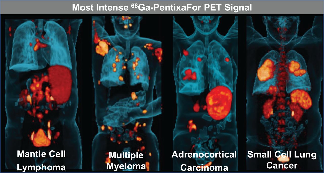
A new molecular imaging radiotracer can precisely diagnose a variety of cancers, providing a roadmap to identify patients who may benefit from targeted radionuclide therapies. In the largest medical study of its kind, researchers found that 68Ga-PentixaFor demonstrated high image contrast in hematologic malignancies, small cell lung cancer, and adrenocortical neoplasms. This research was published in the November issue of The Journal of Nuclear Medicine.
The imaging agent 68Ga-PentixaFor is used to detect the C-X-C motif chemokine receptor 4 (CXCR4). Chemokine receptor expression is a predictor of poor prognosis in cancer patients, as it promotes the growth of malignant cells and inhibits anti-tumor response.
“Knowing which cancers have increased CXCR4 expression can significantly impact clinical tumor staging and, subsequently, affect patient management and treatment decisions,” said Andreas Buck, director of the department of nuclear medicine at University Hospital Würzburg in Würzburg, Germany. “68Ga-PentixaFor PET is of particular interest because it offers improved lesion detection rates over other imaging modalities, such as MRI, CT, and even FDG PET.”
In the study, researchers assessed 68Ga-PentixaFor uptake and image contrast to determine the most relevant clinical applications of CXCR4-directed imaging. Two study sites performed 68Ga-PentixaFor PET on 690 patients with 35 different types of cancer. Images were interpreted and statistical analyses performed.
Very high tracer uptake was found in patients with multiple myeloma, adrenocortical carcinoma, mantle cell lymphoma, adrenocortical adenoma, and small cell lung cancer. Comparable results were recorded for image contrast.
“These results suggest clinical scenarios in which 68Ga-PentixaFor PET may prove beneficial for directing CXCR4-targeted therapies,” said Buck. “This is of particular importance for malignancies that lack a more suitable imaging modality. A pivotal phase III study is planned to continue this research in the second quarter of 2023.”

Graphical Abstract: Most intense 68Ga-PentixaFor PET signal.
The authors of “Imaging of C-X-C Motif Chemokine Receptor 4 Expression in 690 Patients with Solid or Hematologic Neoplasms using 68Ga-PentixaFor PET” include Andreas K. Buck, Niklas Dreher, Alessandro Lambertini, Andreas Schirbel, Samuel Samnick, and Sebastian E. Serfling, Department of Nuclear Medicine, University Hospital Würzburg, Germany; Alexander Haug and Marcus Hacker, Division of Nuclear Medicine, Medical University of Vienna, Vienna, Austria; Takahiro Higuchi, Department of Nuclear Medicine, University Hospital Würzburg, Germany, and Okayama University Graduate School of Medicine, Dentistry and Pharmaceutical Sciences, Okayama, Japan; Constantin Lapa, Nuclear Medicine, Medical Faculty, University of Augsburg, Germany; Alexander Weich, Department of Internal Medicine II, Gastroenterology and ENETS Center for Excellence, University Hospital Würzburg, Germany; Martin G. Pomper, The Russell H. Morgan Department of Radiology and Radiological Sciences, Johns Hopkins School of Medicine, Baltimore, Maryland; Hans-Jürgen Wester, Pharmaceutical Radiochemistry, Technische Universität München, Germany; Anja Zehnder, Department of Nuclear Medicine, University Hospital Würzburg, Germany, and Pentixapharm Würzburg, Würzburg, Germany; Verena Pichler, Division of Pharmaceutical Chemistry, Medical University of Vienna, Vienna, Austria; Stefanie Hahner and Martin Fassnacht, Division of Endocrinology ad Diabetes, Department of Medicine I, University Hospital Würzburg, Würzburg, Germany; Hermann Einsele, Department of Internal Medicine II, Hematology and Oncology, University Hospital Würzburg, Würzburg, Germany; and Rudolf A. Werner, Department of Nuclear Medicine, University Hospital Würzburg, Germany, and The Russell H. Morgan Department of Radiology and Radiological Sciences, Johns Hopkins School of Medicine, Baltimore, Maryland.
