With the benefit of reduced exposure to radiation, PET/MRI could become a practical alternative to PET/CT in the routine management of lymphoma patients, according to a group of Australian researchers.
A team led by Dr. Vijay Mistry of Princess Alexandra Hospital in Brisbane compared the diagnostic performance of same-day FDG-PET/MRI versus FDG-PET/CT in patients with newly diagnosed Hodgkin and non-Hodgkin lymphoma. The approaches matched almost perfectly, yet the group gave PET/MRI the edge given that it exposed patients to less radiation.
"With high concordance in disease site identification, staging, and response assessment, PET/MRI is a potentially viable alternative to PET/CT in lymphoma that minimizes radiation exposure," the team wrote. The study was published online January 24 in Cancer Imaging.
Accurate staging and treatment response assessment are essential for patients with lymphoma. FDG-PET/CT scans are widely used, with FDG radiotracer standardized uptake values providing scores clinicians use to detect changes in tumors. But lymphoma patients diagnosed typically require multiple imaging examinations over the course of diagnosis, treatment, and surveillance. Considering the potential impact of radiation exposure, imaging approaches using MRI are of clinical interest, the authors wrote.
Mistry and colleagues explored the use of a hybrid PET/MRI imaging system in these patients, comparing its performance to FDG-PET/CT scans. They conducted a study that included 50 adult patients with a new diagnosis of any subtype of lymphoma. Patients underwent same-day PET/MRI following PET/CT for staging purposes using the same F-18 FDG injection. All patients were also invited to undergo a PET/MRI after PET/CT scans at interim and end-of-treatment assessments.

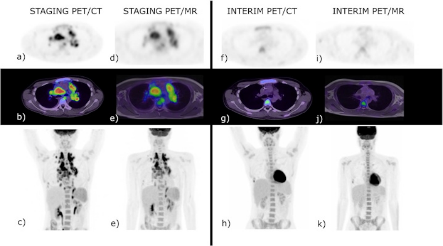
A 20-year-old male with biopsy-proven classical Hodgkin lymphoma, stage IVB. Baseline PET/ CT (a-c) and baseline PET/MR (d-e). At interim restaging PET/CT (f–h) and PET/MR (i-k), there was a concordant complete metabolic response, Deauville 2. The end of treatment study is not shown as there remained complete metabolic response. Incidental right-sided rib fractures persisted between staging and interim studies. SUV scale set between 0-8. Image courtesy of Cancer Imaging through CC BY 4.0.
At staging, PET/CT detected 352 positive disease sites out of a potential 1,500 sites across 50 patients. PET/MRI detected 339 out of 352 true-positive sites, 1,148 true-negative sites, and 13 false-negatives sites. There was excellent intermodality agreement between PET/MRI and PET/CT, the researchers reported (k = 0.976, p < 0.001). There was also good correlation of disease site identification at interim assessment (k = 0.819, p < 0.001) and excellent correlation at end-of-treatment assessment (k = 1, p < 0.001), they reported.
Another benefit of PET/MRI? Lower patient effective dose (ED): ED for PET/CT was 10.5 mSv per scan, while the average calculated ED for the PET/MR was 4.8 mV per scan, resulting in a difference of 5.7 mSv dose decrease (54%) between the two modalities.
"With the benefit of avoiding radiation exposure, PET/MRI appears an attractive alternative to PET/CT across indolent and aggressive lymphomas with comparable performance," the authors wrote.
Ultimately, PET/CT is the current standard in imaging of lymphoma patients due to availability and familiarity. This study supports the introduction of PET/MRI into clinical practice, yet further research on its diagnostic performance is crucial, the team noted.
"Given the contemporary nature of PET/MRI, studies with multiple institutions with standardized imaging protocols would allow further assessment of inter-equipment variability and would benefit from a larger recruitment population," Mistry and colleagues concluded.