PET imaging shows promise in the difficult task of differentiating between subtypes of atypical parkinsonism, according to a study published August 12 in EJNMMI Research.
A team led by Naba Jawad Houssein, of Copenhagen University Hospital Bispebjerg in Copenhagen, Denmark, demonstrated that cerebral F-18 FDG-PET scans helped differentiate between subtypes of atypical parkinsonism with significant sensitivity (> 80%) and specificity (> 90%). The results support the use of PET for the clinical diagnosis of atypical parkinsonism, the group noted.
"Precise [atypical parkinsonism] diagnosis is crucial for accurate prognosis and effective therapy, but the disease is often difficult to diagnose in early stages based on clinical criteria alone," the researchers wrote.
Atypical parkinsonism includes a variety of neurological disorders in which patients share some clinical features of Parkinson's disease. These disorders are distinct, however, and may include multiple system atrophy, progressive supranuclear palsy, corticobasal degeneration, and Lewy body dementia.
While the diagnosis of atypical parkinsonism is based on clinical criteria, the researchers hypothesized that F-18 FDG-PET scans could provide more detailed data to separate the disorders based on uptake of FDG radiotracer in affected brain regions. Moreover, only a few studies have evaluated the sensitivity and specificity of the technique for this use, the researchers added.

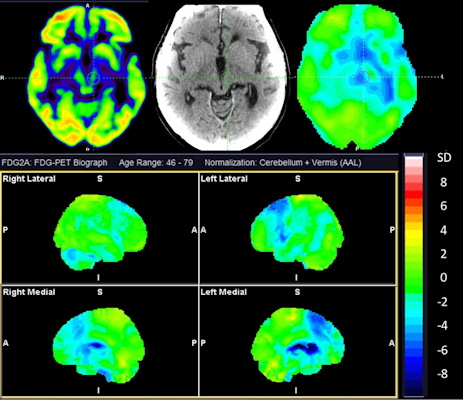
Cerebral F-18 FDG-PET of a 73-year-old man with overlapping features of corticobasal degeneration (CBD) and progressive supranuclear palsy (PSP). The top row shows axial sections at the level of the basal ganglia and mesencephalon of F-18 FDG, CT, and statistical maps. The bottom rows show statistical surface projections with standard deviations from healthy subjects. Note the asymmetry and involvement of the basal ganglia pointing towards CBD and the involvement of mesencephalon and mesial frontal cortex pointing towards PSP. Image and caption courtesy of EJNMMI Research through CC BY 4.0.
The group identified 156 patients referred for a brain F-18 FDG PET scans between 2017 and 2019 for suspicion of atypical parkinsonism, with imaging analyzed by a nuclear medicine specialist with more than 10 years of experience in PET neuroimaging. The reference standard for comparison was each patient's follow-up clinical diagnosis.
The overall accuracy for correct classification was 74%, according to the results, with sensitivity and specificity for specific disorders as follows:
Multiple system atrophy (n = 20): 100% and 91%
Lewy body dementia (n = 26): 81% and 97%
Progressive supranuclear palsy/corticobasal degeneration (n = 68): 62% and 97%
One of the strengths of the study was that it included a diverse patient population consecutively referred for cerebral F-18 FDG-PET imaging due to suspected atypical parkinsonism, and thus reflects real-world clinical practice, the authors wrote.
They also noted that promising radiotracers such as F-18 APN-1607 and F-18 PI-2620 have been developed to provide even more specific detail on atypical parkinsonism subtypes, and with further study could greatly aid in early diagnosis.
"Use of F-18 FDG-PET may be beneficial for prognosis and supportive treatment of the patients and useful for future clinical treatment trials," the researchers concluded.