Flortaucipir PET imaging is able to identify neuropathological brain changes in retired contact sport athletes at risk of neurodegeneration, according to a study published July 22 in the Journal of Neurology.
A group at the University of Toronto in Ontario, Canada, compared flortaucipir (Tauvid, Avid Pharmaceuticals) PET and MRI scans between retired athletes who experienced concussions and normal controls. The team found that a higher tau-PET signal was associated with reduced grey matter volumes and lower memory scores.
“Tau PET may be useful for identifying those at risk for neurodegeneration,” noted first author Anna Vasilevskaya, PhD, of the university’s Tanz Centre for Research in Neurodegenerative Diseases.
Concussions and mild traumatic brain injuries have been linked to tau brain pathology and associated with neurodegeneration in both Alzheimer’s disease and chronic traumatic encephalopathy (CTE) patients, the authors explained.
However, while flortaucipir PET scans are approved in the U.S. and widely used for identifying patients with suspected Alzheimer’s disease, the utility of the scans for detecting tau pathology in CTE is still unclear, they noted. Thus, the group’s main objective was to evaluate flortaucipir PET imaging findings in a cohort of retired contact sport athletes at risk for CTE due to concussions, yet who were negative for Alzheimer’s disease biomarkers.
The researchers enrolled 47 retired contact sport athletes negative for Alzheimer’s disease (mean age: 51 ± 14; concussions per athlete: 15 ± 2) and 54 normal controls (mean age: 50 ± 13) through the Canadian Football League Alumni Association or the Canadian Concussion Center clinic in Toronto. Participants underwent MRI, flortaucipir PET, and a neuropsychological battery.
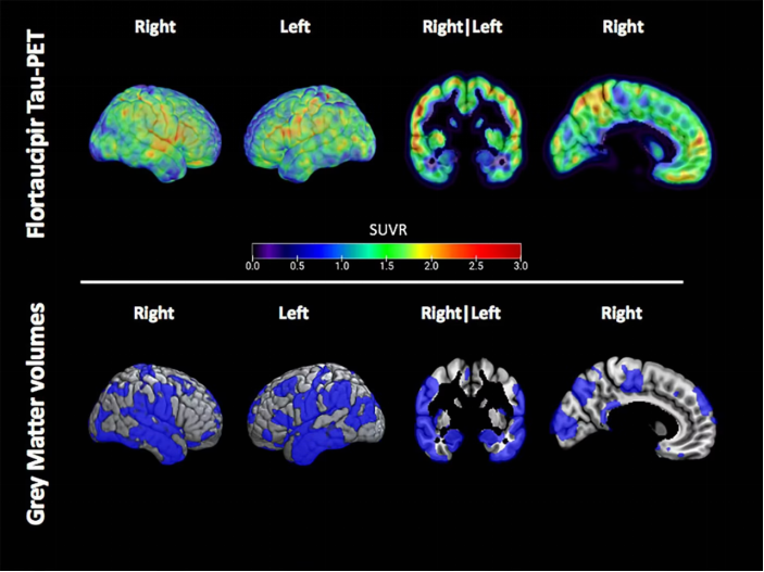
Example of the flortaucipir tau-PET signal and distribution of low grey matter volume in a single former athlete. Spatial maps of the flortaucipir tau-PET standardized update value (SUVR) (top) and the reduced grey matter volumes showed in blue and defined as voxels with a w-score of >1.5 below the mean (bottom) in a single former athlete. Image courtesy of the Journal of Neurology.
According to the analysis, tau-positive voxels on PET scans also had significantly lower gray matter volumes on MRI in the athletes, compared with tau-PET negative voxels. In addition, there was a significant relationship between these tau-PET positive measurements and patients who had lower memory scores.
“We found that tau-PET positive regions had lower grey matter volumes than did the tau PET negative regions, suggesting an underlying pathophysiological process causing grey matter atrophy,” the group wrote.
Notably, the spatial pattern of the tau PET signal involved mostly frontal and temporal brain regions and extended posteriorly as levels increased, which is consistent with postmortem studies that reported tau pathology in patients with CTE, the authors wrote.
There was no relationship between tau PET measures and concussion number, or years of sport played, they added.
Ultimately, the meaning of flortaucipir radiotracer binding in former contact sports athletes and its ability to reflect CTE has been a matter of debate, the authors noted. This study supports evidence that flortaucipir PET is able to identify neuropathological changes in retired athletes at risk of neurodegeneration, they wrote.
“However, more studies examining the relationship between flortaucipir tau-PET and postmortem CTE pathology are needed to identify its specificity to CTE pathology,” the group concluded.