PET/MRI breast cancer imaging time can be cut in half without losing key diagnostic information -- a finding that could increase the use of the modality for this indication and streamline radiology department throughput, researchers have reported.
First author doctoral candidate Kai Jannusch of the University of Dusseldorf and colleagues shortened F-18 FDG-PET/MRI protocols from 20 to under 10 minutes in scans of breast cancer patients with small tumors without sacrificing essential findings, according to a study published April 12 in European Radiology.
"[F-18 FDG-PET/MRI] is of great importance due to its local tumor staging and phenotyping abilities and should not be skipped aiming towards faster examination protocols," the group noted. "Reducing the time of PET data acquisition while implementing a shortened but still diagnostic breast MRI protocol might solve the problem of long examination times."
Accurate staging is crucial in women after initial diagnoses with breast cancer, and PET/MRI has begun to emerge as a technique in these cases for local and whole-body staging. But a well-known barrier to increasing the use of hybrid exams is the time they take.
"A key problem of F-18 FDG-PET/MRI breast cancer staging is the time-consuming nature," the group wrote. "With a 20-minute acquisition time, the dedicated breast F-18 FDG-PET/MRI is a time-consuming part of the overall examination process."
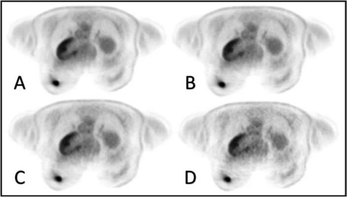
To determine if they could speed the exams up, the researchers conducted a study that included 90 women with newly diagnosed T1 and T2 breast cancer who underwent breast F-18 FDG-PET/MRI scans, then used software to reconstruct the imaging data and simulate PET acquisition times at 20, 15, 10, and five minutes. Nuclear medicine and breast imaging radiologists then analyzed and compared the images in random order, assessing image quality.

PET images of a 54-year-old breast cancer patient with a good subjective image quality score (5-point scale: 4) for all reconstruction times of 20 minutes (A), 15 minutes (B), 10 minutes (C), and five minutes (D). Image and caption courtesy of European Radiology through CC BY 4.0.
In all of the PET reconstructions, the readers detected 127 congruent breast lesions. Comparisons among T1 versus T2 subgroups showed no significant differences in subjective image quality between the 20-, 15-, 10-, and five-minute acquisition times, according to the investigators.
Specifically, the group found that breast F-18 FDG-PET/MRI protocols could be shortened from 20 to approximately eight minutes without losing essential diagnostic information.
"This enables a considerable reduction of the breast F-18 FDG-PET/MRI protocol and consequently higher patient throughput combined with greater patient satisfaction," the group wrote.
This study is the first to evaluate fast breast F-18 FDG-PET/MRI scans in this patient group, according to the researchers, but further studies are warranted.
"Aiming towards faster breast F-18 FDG-PET/MRI protocols, this is the first study that systematically investigates the effect of reduced PET acquisition times on PET image quality and quantification parameters as well as a shortened, fast breast MRI protocol in a clinical setting of T1 and T2 breast cancer," the group concluded.