Every year, there are more than 4 million new cancer patients in China, among which more than 1.2 million patients are at great risk of misdiagnosis, mistreatment and over-treatment, since they can't get accurate diagnosis. Because of its complex pathogenesis and huge individual differences, the current treatment efficiency of tumors is very low. Even if high-efficiency anticancer drugs and mature immunotherapy are developed, everything is empty talk without accurate diagnosis of tumor types. Accurate diagnosis of tumors is the prerequisite to defeat tumors.
With the development of new imaging technologies, especially the introduction of PET/CT (Positron emission tomography and CT integrated machine) and PET/MR (Positron emission tomography and MRI integrated machine), nuclear medicine has excellent ability of lesion detection and analysis, playing an increasingly important role in the accurate diagnosis of major diseases such as tumors.
18F-FES
18F-Fluoroestradiol(FES) is an estrogen analogue, which has been proved to be a promising biomarker in ER imaging of breast cancer. FES uptake is related to ER expression, which can be used to qualitatively and quantitatively evaluate multiple tumor sites and predict the response of endocrine therapy.
18F-FDG
2-deoxy-2-[fluoro-18] fluoro-D-glucose (18F-FDG) contained in the positron emission tomography (PET) is an analogue of glucose, which provides valuable functional information based on the increase of glucose uptake and glycolysis in cancer cells, and describes the metabolic abnormality before morphological changes. 18F-FDG PET/CT is more sensitive and specific in some cancers, and it is mainly used as a staging and re-staging tool to guide patient care.
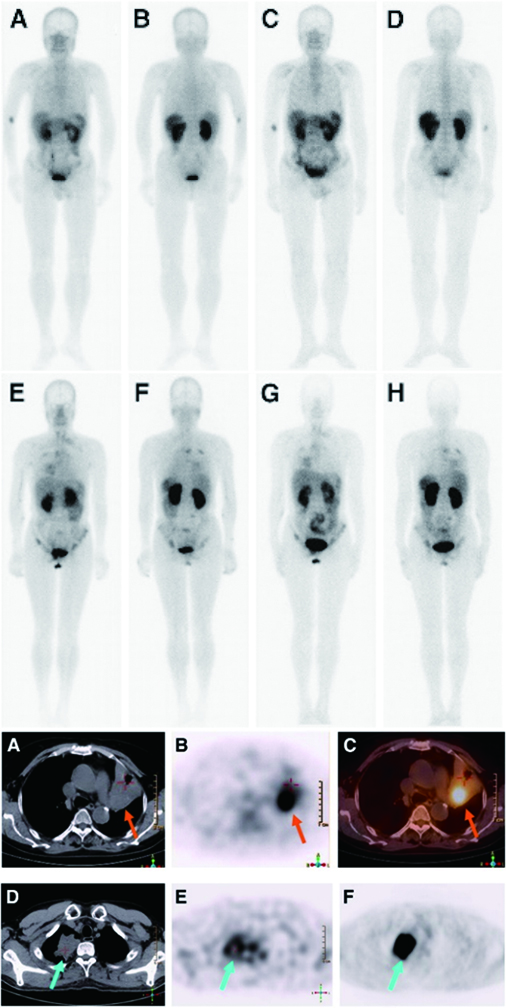
Accurate diagnosis of metastatic/primary cancer
CT imaging (B, D) showed an irregular mass in the left lung with a maximum diameter of 4.5 cm of the patient. It is difficult to distinguish metastatic lung cancer from secondary primary lung cancer on CT.
Using radiopharmaceutical PET/CT imaging (A.18F-FES-PET/CT, C.18F-FDG-PET/CT), the tumor showed high uptake of 18F-FES and 18F-FDG PET, and the SUVmax was 6.3 and 5.5, respectively. The results suggested that the tumor of this patient was metastatic tumor.
ER positive metastasis of breast cancer was confirmed after operation.
Journal Title:
Sun Y, Yang Z, Zhang Y, Xue J, Wang M, Shi W, Zhu B, Hu S, Yao Z, Pan H, Zhang Y. The preliminary study of 16α-[18F]fluoroestradiol PET/CT in assisting the individualized treatment decisions of breast cancer patients. PLoS One. 2015 Jan 24;10(1):e0116341.
68Ga DOTATOC was approved by the U.S. Food and Drug Administration (FDA) in 2019 as the first 68Ga radiopharmaceutical for imaging somatostatin receptor (SSTR)-positive gastrointestinal and pancreatic neuroendocrine tumors using positron emission tomography (PET). This radiopharmaceutical combines radionuclide 68Ga with somatostatin analog DOTA-TOC to specifically image tumor cells expressing SSTR. This targeting method can also be used for treatment planning of local and disseminated diseases, as well as for evaluation of treatment response.
68Ga DOTATOC PET/CT detection
68Ga DOTATOC PET/CT provides one-stop multi-site accurate diagnosis of the whole body, including evaluation of liver, lymph nodes, bone, lung, brain and other possible tumor sites.
In the figure, 68Ga DOTATOC accurately shows the patient's ileum (B) primary neuroendocrine tumor with lymph node (C) and bone metastasis (D)
Journal Title: Theranostics. 2012; 2(5): 437–447.
The integrin alpha(α)v beta(β)3 receptor is ubiquitous in malignant tumors and has a certain level of specificity for tumors. Technetium-99m hydrazinonicotinamide-dimeric cyclic arginyl-glycyl-aspartic acid peptide with three polyethylene glycol spacers (99mTc-3PRGD2) can bind specifically to the integrin αvβ3 receptor with high selectivity and strong affinity. Thus, it can specifically mark tumors and regions with angiogenesis for tumor detection and be used in single-photon emission computed tomography (SPECT) imaging.
99mTc-3PRGD2 SPECT imaging can be applied to the diagnosis of tumors positive for the integrin αvβ3 receptor, determination of the location and extent of tumors and metastases, monitoring of postoperative residual or recurrent lesions, auxiliary diagnosis of neovascular density, and efficacy and prognostic evaluation of tumor anti-neovascular therapy.
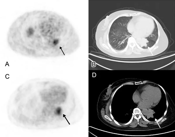
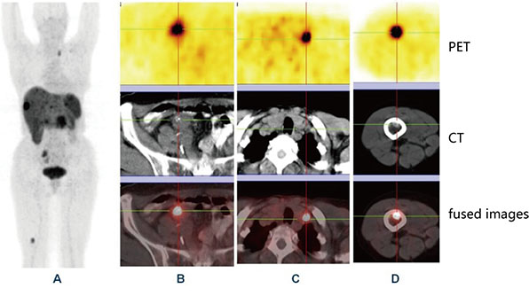
99mTc-3PRGD2 for Integrin Receptor Imaging of Lung Cancer
With a low 99mTc-3PRGD2 background in the lungs and mediastinum, most lung malignancies were prominent on 1-h images with tumor-to-background ratios significantly higher than those in benign lesions. Furthermore, most lymph-node and bone metastases could also be detected. Thus, the findings suggested that 99mTc-3PRGD2 SPECT imaging is a sensitive tool for detecting lung cancer (sensitivity, 93–97%).
Journal Title:
99mTc-3PRGD2 for Integrin Receptor Imaging of Lung Cancer: A Multicenter Study. J Nucl Med (2012) 53(5):716–22.
Comparison of the Accuracy of 99mTc-3P4-RGD2 SPECT and CT in Diagnosing Solitary Pulmonary Nodules. Oncol Lett (2016) 12(4):2517–23